
3D Mammography Improves Detection, Reduces Call Backs
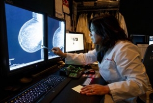
Radiologist Debra Ikeda, MD, reviews a breast image created with Stanford’s new 3D tomosynthesis system and C-View software. 3D tomosynthesis improves detection of small cancers, and reduces call backs.
With no magic bullet or blood test to detect breast cancer, radiologists like Debra Ikeda, MD, are always investigating new technologies to improve early detection of tumors. Ikeda first saw 3D tomosynthesis in 2006, and she’s been waiting for FDA approval in the U.S. to bring it to Stanford. Beginning in April of this year, Stanford began offering 3D tomosynthesis to all screening patients at its two breast imaging centers in Palo Alto.
"The problem women face in mammography is that a small cancer can be hidden by breast tissue overlying or underlying it," says Ikeda, a professor of radiology who directs Stanford’s Breast Imaging Section. "With 3D tomosynthesis, we’ll be able to see a finding that was previously hidden."
In numerous research studies conducted in Europe, 3D tomosynthesis in combination with 2D mammography provides about a 25 percent improvement in overall cancer detection rates, and a 15 percent reduction in false positives. This greatly reduces the number of women who have to be called back for secondary testing, says Ikeda. Further studies were conducted on the use of 3D tomosynthesis alone, with C-View software to create the 2D image, and the results were equally promising.
Now we can find many little cancers that were being obscured by dense tissue.
How it works
3D mammography can be done in conjunction with a traditional 2D digital mammogram, or separately using the C-View synthesized software. During the 3D part of the exam, an X-ray arm sweeps in a slight arc over a patient’s breast, taking multiple breast images. Then, a computer produces a 3D image of the breast tissue in slices, like a CT Scan, providing greater visibility for the radiologist to see breast detail in a way never before possible. The C-View software is used to create a synthesized 2D image from tomosynthesis data sets, eliminating the need for a separate 2D exposure.
3D mammography complements standard 2D mammography and is performed at the same time with the same system. There is no additional compression required, and it only takes a few more seconds longer for each view. The radiation dose with tomosynthesis and C-View synthesized 2D software is about the same average dose of 2D digital mammograms in the USA.
"With synthesized view, you take all the images in 3D slices and then use the computer software to reconstruct an image of the entire breast without taking another picture," says Ikeda. By looking at breast tissue in one-millimeter slices, 3D mammography can show cancers missed by conventional 2D mammography. This allows the radiologist to provide a more confident assessment, says Ikeda.
Who receives 3D tomo?
Small breast cancers can go undetected in women with dense breast tissue, as well as in women with less dense breasts, says Ikeda. So every woman who comes to Stanford for screening will be offered 3D tomosynthesis. Selected diagnostic patients will be offered 3D tomosynthesis based on the woman’s problem.
"3D is especially good at finding small, stellate cancers," she says, "and it will be really helpful in finding cancers in women undergoing screening, or second tumors in women who already cancer."
But Ikeda cautions that tomosynthesis still misses some cancers, especially round tumors hidden among dense tissue. Finding those types of cancer will take a different type of technology such as MRI, or contrast-enhanced mammography which Stanford plans to add next year.
By Grace Hammerstrom