
Stanford Implants First Responsive Neurostimulator to Treat Intractable Epilepsy
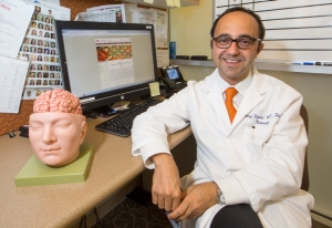
Josef Parvizi, MD, director of Stanford's Program for Intractable Epilepsy celebrates the first two implantations of the new NeuroPace responsive neurostimulator system in patients with intractable epilepsy.
Stanford Health Care is one of the first hospitals in the country to implant the NeuroPace responsive neurostimulator system (RNS) in patients following FDA approval of the device last fall. Embedded completely within the skull, the device monitors electrical activity in the brain, senses abnormal activity and responds by delivering unnoticeable pulses of electrical stimulation to normalize that activity before a person experiences a seizure.
Candidates for the RNS device include patients who suffer multiple seizures a day, who are on two or more antiepileptic drugs that no longer work effectively and whose seizures originate in one or two localized areas of the brain.
"This device is going to be used mainly for patients who have seizures in eloquent brain—the speech and primary motor and sensory areas—where a surgical resection would leave them with a deficit that might not be acceptable," says Michael Edwards, MD, Lucile Packard Endowed Professor of Neurosurgery, who performed the first RNS implantation at SHC.
"It truly expands the number of patients we can treat and help reduce their epilepsy," says Jaimie Henderson, MD, the John and Jene Blume–Robert and Ruth Halperin Professor of Neurosurgery, who just a week later, performed the second RNS implantation surgery at Stanford. "There are quite a few patients who are not candidates for traditional epilepsy surgery and who have failed medical treatment. They may now have this as an option."
How it works
The battery-powered and microprocessor-controlled device is placed within the skull and beneath the scalp. It is connected to one or two leads containing electrodes that are placed within the brain in the area of the seizure focus. Patients can be treated with one device to stimulate one or two areas of the brain, or two devices to stimulate up to four areas of the brain.
After the RNS system is implanted, it immediately begins recording seizure activity in the brain. The device learns to sense the onset of a seizure, and then triggers an electrical impulse back down through the electrodes to silence or quiet that area, says Edwards. Physicians can program the detection and stimulation parameters of the implanted RNS system non-invasively to optimize therapy for each individual.
Pioneering patients
The first patient treated at Stanford with the RNS system was considered a candidate for the device only after it was determined that resection surgery would impair his leg function, says Josef Parvizi, MD, PhD, associate professor of neurology and director of Stanford's Program for Intractable Epilepsy. This patient's seizures were emanating from the sensory motor area of the brain. A resection could have eliminated his seizures, says Parvizi, but it would have also had consequences such as foot weakness, numbness or even paralysis. "This was not a risk this young, active patient was willing to take," he adds.
The second patient, a young woman with temporal lobe epilepsy, was found to have bilateral seizure foci in both of her hiccocampi. Although you can often remove one hippocampus with little deficit to the patient, says Henderson, removing both hippocampi would remove the patient's ability to form new memories. "For her, the RNS was really the only option that would allow us to potentially treat her seizures," he says.
With one million people in the U.S. with intractable epilepsy, there are many patients who may be candidates for this type of treatment, says Parvizi, including patients who were previously not considered candidates for resective surgery. "Even though it's an invasive procedure, implanting a device into a patient's brain, the RNS system has the advantage of not destroying the brain."
Isolating the trouble spot
Not all seizures are suitable for treatment by responsive neurostimulation. First, the location of the seizures in the brain must be pinpointed, and second, the seizures must be localized in one or two spots. All patients evaluated for epilepsy surgery at Stanford undergo combined seizure and functional mapping to isolate the location of the seizure focus, and determine the risk of resecting in a specific area.
To do this, neurosurgeons and neurologists work together to implant electrodes over areas of the brain where they believe the seizures are originating, and monitor seizure activity for four to five days. Through video and EEG monitoring, we can determine where the seizures start, says Edwards. These electrodes can then be used to stimulate brain tissue to identify the underlying function of the region, such as language, sensation or motor function. This functional mapping helps surgeons avoid critical areas while operating.
The NeuroPace RNS system has been evaluated in three clinical trials, and demonstrated a 37.9 percent reduction in seizure frequency. For those patients who reached two years post implant, 55 percent experienced a 50 percent or greater reduction in seizures. When Edwards looked further at the data on patients implanted in the sensory motor area, he learned that 80 percent had complete resolution of their seizures with minimal medicine or no medicine at all. "That," he says, "is an amazing response."