
Endovascular Treatment Improves Outcomes for Ischemic Stroke
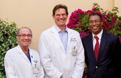
(Left to right): Michael Marks, MD, Greg Albers, MD and Robert Dodd, MD, PhD, collaborate to bring the latest standard of care for stroke – endovascular therapy – to patients at Stanford.
With stroke, time is still essential to preserving brain tissue. But new endovascular clot retrievers are improving outcomes, reducing mortality and extending the treatment window for many patients suffering an acute ischemic stroke. In addition, advanced imaging techniques have revealed that many patients have salvageable brain tissue well beyond the traditional three-hour treatment window, allowing more patients to be treated successfully.
"The outcomes for stroke have never been better," says Greg Albers, MD, director of the Stanford Stroke Center. In four recently reported studies, endovascular clot retrieval has been found to significantly improve patient outcomes from ischemic stroke. "Endovascular therapy has become a new standard of care for stroke," adds Albers. "It is now completely and thoroughly proven that appropriately selected patients will benefit from this procedure within six hours of symptom onset."
Improved retrievers transform outcomes
The most common type of stroke, an ischemic stroke, occurs when a blood clot goes up into the brain and blocks off the blood flow. With 700,000 ischemic strokes in the U.S. alone, it's the number one cause of disability and a huge public health problem.
For the past 20 years, clinicians have relied on one treatment, clot-dissolving medicine called TPA to return blood flow to the brain, a process called reperfusion. But TPA must be administered within three-hours of having a stroke for the greatest chance of success. Even then, it is only successful in about one third of patients. Researchers at Stanford and other academic medical centers across the U.S have been studying the benefits of removing clots via endovascular techniques, opening up clogged arteries and bringing oxygenated blood to starved brain tissue.
"The question has been, can endovascular surgeons do better than TPA by physically pulling the clot out?" asks Albers, the Coyote Foundation Professor of Neurology, and Professor by Courtesy of Neurosurgery. By threading a catheter through a blood vessel in the leg, up into the brain, endovascular surgeons can locate the clot and pull it out. Up until very recently, it was uncertain whether the procedure was actually making patients better, partly because the original devices were not extremely effective at removing clots in their entirety.
But improved devices have transformed outcomes. Called stent retrievers, these catheters do a much better job at getting vessels open than earlier generation devices. "These newer devices have led to phenomenally better reperfusion rates than we were getting with our old devices," says Michael P. Marks, MD, professor of radiology and by courtesy of neurosurgery. "When we used older devices, we were able to get the brain reperfused in a way that we felt was going to be beneficial only about 40 percent of the time," he adds. "Now we’re approaching 80 to 90 percent."
Imaging guides care
In parallel with needing better devices, clinicians needed better imaging to determine which patients would benefit from reperfusion. Over the past 15 years, Stanford has pioneered several imaging techniques including MRI techniques and CT perfusion that facilitate the identification of salvageable ischemic brain tissue in patients presenting with an acute stroke.
The speed at which brain cells die post stroke is highly variable. In some patients, brain cells die very quickly, even within the three-hour window commonly used to treat stroke. Others have much more resilient tissue that is salvageable hours after a stroke. The difference lies in what's known in the stroke world as collaterals. Patients who experience rapid brain tissue death do not have adaquate blood vessels nearby to provide blood flow to the area affected by the stroke. Good collaterals give patients a better chance of limiting the amount of brain tissue that ultimately dies.
"The blood vessels supplying our brains were designed to allow for blood flow to bypass a blockage, at least for a temporary time, and get supply from other large vessels at the base of the brain," says Marks. "These are the collateral supply of blood flow."
By using advanced imaging techniques, and processing them in such a way as to get maps of what the brain looks like, we can see the tissue that is already damaged and the tissue that is able to be salvaged, and we can get a very good picture of how much we can help a patient by opening up that circulation, he adds.
In a recent case, Robert Dodd, MD, PhD, assistant professor of neurosurgery and radiology, treated a 91 year old woman from Eureka who arrived at Stanford nine hours after her stroke onset, outside the standard window of treatment for either medical or endovascular therapy. Perfusion imaging revealed that she still had a sizeable amount of salvageable tissue, so Dodd removed her clot endovascularly and she recovered well.
Not all patients are so lucky. For those with insufficient collateral blood supplies and rapidly dying brain tissue, there is little benefit derived from clot dissolution or removal. But many, those with good collaterals and a low percentage of dead tissue, can be treated successfully even when they arrive at the hospital outside of the three-hour window. This is good news for patients, says Albers, because more than half of patients with ischemic stroke are getting to the hospital outside of the six-hour window. "Imaging lets us know if the patient can be helped, regardless of when they arrive," adds Marks.
"The current revolution in stroke care, which includes endovascular therapy, perfusion-based imaging and neuro-critical care, has now clearly demonstrated to overwhelmingly improve outcomes," says Dodd.