
Microvascular Reconstruction Improves Results for Head and Neck Cancers
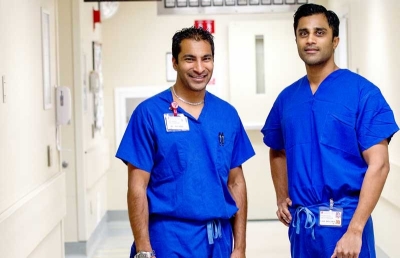
Left to right: Rahim Nazerali, MD, in the Division of Plastic and Reconstructive Surgery, and Vasu Divi, MD, in the Division of Otolaryngology - Head & Neck Surgery, often collaborate on cancer resection and reconstruction surgeries that require advanced microvascular techniques to rebuild tongues and jaws lost to cancer.
With microvascular reconstruction, surgeons move a composite piece of tissue from one part of the body to the head and neck to rebuild jaws, optimize tongue function and reconstruct the throat. Although microvascular reconstruction itself is not a new field, it has vastly improved over the past 40 years when success rates were low and the rate of flap loss was very high. Today, surgeons have a much better toolbox with which to work, and are able to achieve excellent reconstructive, aesthetic and functional results.
"We're able to isolate blood vessels in the body that are associated with either skin, fat, muscle or bone," says Rahim Nazerali, MD, clinical assistant professor of surgery in the Division of Plastic and Reconstructive Surgery. "We're able to detach those structures on their blood vessels and then auto transplant them to another location on the body to help restore form and function."
Bringing along the blood vessels with the transplanted tissue is essential to keeping the tissue alive in its new home, says Vasu Divi, MD, assistant professor, Otolaryngology - Head & Neck Surgery. "I tell patients it's the difference between laying down sod and moving a tree. The tree needs to come with its own roots."
With improved knowledge of anatomy, improved microscopes and microsurgical tools and improved techniques, surgeons such as Divi, Nazerali and their colleagues are able to achieve successful outcomes 95 to 98 percent of the time, removing cancers today that at one time were unresectable.
Tongue Reconstruction
In 2012, Sherri Murphy was diagnosed with Stage 3 tongue cancer affecting more than half of her tongue. When her physician told her surgery was not an option, Murphy came to Stanford for a second opinion. "Stanford was my life-saver," she says. "I learned surgical resection was a possibility."
In the four-hour reconstruction surgery, Divi took a section of Murphy's forearm, along with its blood vessels intact, and transplanted it to the remaining portion of her tongue. In the past, surgeons relied on thin skin grafts to reconstruct tongues, he says. This left patients' tongues stuck to the bottom of their mouths, severely impairing their speech and ability to eat.
Tongues require mobility and volume to be functional. They must have enough volume to touch the roof of the mouth and the back of the front teeth to obliterate the oral cavity when a person swallows. Tongue reconstruction using a thick piece of patient's forearm provides the volume necessary to allow the remaining section of the tongue to do its function.
"My goal with tongue resections is to get patients back to speaking in an understandable way," says Divi. "If they can talk to someone over the phone and be understood, that's the best. To be understood with no verbal cues, no lip reading and no hand gesturing, that to me is the highest level at which we operate."
Kimberley Garrett is just such a patient. A customer service supervisor for Verizon, Garrett spends much of her day on the phone. For her, speaking clearly is a necessity. Three years ago, at age 23, she was diagnosed with stage 4, squamous cell carcinoma on her tongue. In a 12-hour surgery, Divi removed half of her tongue and rebuilt it using a flap of skin from her forearm. Although her recovery was long, today, her speech is flawless.
"When I have to remove half or less than half of a patient's tongue, I am very confident about what we can do to get the patient back to speaking and eating," says Divi. Patients who lose one half to three fourths of their tongue have more variable results, he says.
3D Jaw Reconstruction
Whether a patient loses bone in the jaw due to cancer resection or radiation-related damage, the same principles of transplantation apply. But rather than moving muscle, surgeons must move bone. Ensuring that this bone fits precisely into the hole left in the patient's jaw requires a multi-step process using 3-D simulation and modeling.
The first step in a jaw resection and reconstruction, says Divi, is to create a precise 3-D model from a patient's CT scans. This model is created at a specialized center in Michigan, and is available to the surgical team via remote link. Viewing the patient's jaw in computer simulation, the surgeon can precisely mark out where to remove the bone, and create a mirror image on the opposite side of the jaw to fill in the gap.
"That's how we achieve a symmetric jaw with everything in the correct position," he says. Once the resection and reconstruction is mapped out, Divi requests a 3-D print, a plastic model of the patient's jaw, which he uses to guide the reconstruction by bending flexible metal plates along the model's jawline to capture its precise contours.
"Ultimately what I need going into the OR is a metal plate that spans the gap left behind from the cancer, that's custom designed to that patient's jaw and that keeps the teeth perfectly aligned," he says. During surgery, he screws this plate onto the patient's jaw to make sure the teeth line up in the same position afterwards. He then removes the plate, removes the diseased bone and then repositions the plate onto the jaw using the pre-drilled holes to guide him as he fills the gap with the transplanted fibula.
"The plate is the framework," says Divi. "But getting the plate right takes a lot of planning."