
Meniscus Tear
The knee has two cartilage cushions that are situated between the femur (thigh bone) and tibia (shin bone) that function as shock absorbers, force distributors, and secondary restraints to abnormal motion. These are called menisci (plural for meniscus). They are in the configuration of 2 half circles, one medial (inside part of the knee) and one lateral (outside part of the knee), and face each other. They have attachments to the surrounding joint capsule, the MCL, and the tibia to help maintain them in the correct position as the knee moves.
The primary role of these fibrocartilage rings are to attempt to evenly distribute the force of the body weight with such activities as walking, running and jumping such that the articular cartilage (cartilage lining the ends of the bones) is protected from excessive force.
Unfortunately, these very important structures in the knee are also very prone to injury. This is based on their shape or configuration and their limited blood supply (which is required for healing). The tissue itself also slowly degenerates over time as we age and makes them even more prone to damage. This leads to "degenerative tearing" where the tissue starts to breakdown and weaken forming complex tears that are typically not repairable. Most tears in the mensicus have a very limited chance of healing based on how the flow of blood is directed. However, those tears that occur in young and healthy individuals in a specific location of the meniscus and are treated early, can often be surgically repaired.
Injury occurs typically from a twisting motion in a knee that is flexed as this piece of cartilage is trapped between the two bones of the knee. These are also commonly injured when ligaments are torn in the knee as well. A meniscus tear will typically present with intermittent pain in the knee which can be isolated to one side and may be both dull and achy as well as very sharp. This is usually worse with deep knee flexion (squatting motions) or with rotation or pivoting. Often, the knee will become swollen and a clicking or catching sensation may also be experienced. Occasionally the knee will actually lock (become stuck) during routine motion or walking which can be very painful, debilitating, and is potentially a sign of a significant problem that may require surgery.
The diagnosis of a meniscus tear is made based on the presenting complaint, the mechanism or onset of pain, a careful physical examination of the knee and X-rays and an MRI. Several physical exam tests have been described by orthopaedic surgeons and when all of them are combined, they are quite reliable for making the diagnosis. The X-rays are used to identify the mechanical alignment of the knee, any underlying arthritic changes, and a variety of other associated conditions that could affect the prognosis. The MRI is a key item for documenting the location, configuration and extent of the tear. Although not 100% accurate, the MRI helps give the orthopaedic surgeon a better chance at providing a prognosis for any surgical treatment, as well as what special techniques may be required during the surgery.
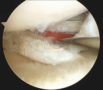
Typically the surgical intervention of choice for a mensicus tear is a knee arthroscopy, which has become one of the most common surgical procedures in orthopaedics. However, in recent years we have learned a tremendous amount about the repair techniques and ways to augment healing of the meniscus. Occasionally, a tear will require an additional procedure paired with the arthroscopy to perform a more extensive repair to maximize the chance of healing the injury. Most commonly, the meniscus tear is not repaired and the torn or loose portion is removed during arthroscopy. The minimum amount of meniscus is removed during this surgery to alleviate symptoms while preserving the functionality of the remaining meniscus and the long term health of the knee.
While the surgery may take the same amount of time for a repair or a partial removal of the mensicus, the rehabilitation process is quite different and this should be discussed with your surgeon.
Meniscus Tear
Two cartilage menisci are within the knee joint allowing smooth movement of the bones, against each other. A meniscus tear causes a lack of stability and discomfort.
Meniscus Tear
meniscus-tear