Osteoid Osteoma Treatment with MR Guided Focused Ultrasound (MRgFUS)
Although osteoid osteomas may regress spontaneously over the course of years, current standard of care in patients whose pain is not controlled by medical therapy is a minimally invasive treatment with radiofrequency ablation.
Under CT guidance, a thin drill needle is used to place a cannula into the bone at the tumor focus. The cannula is used to position a radiofrequency electrode, which is then used to heat the tumor to the point that the tumor around the tip of the electrode is coagulated. After radiofrequency ablation, the tumor pain resolves within a few days, and treatment related pain resolves within a few days to a week. Depending on the location of the tumor, weight-bearing activities may be limited for up to 3 months after treatment. The examples below show treatment of osteoid osteomas in the right femur (A) and right tibia (B) with radiofrequency ablation.
The current main alternative treatment approach is surgical resection. After surgery, patients may spend up to a week in the hospital, and may require up to 6 months before they can return to normal activities. Sometimes surgery requires prophylactic placement of internal fixation hardware, if resection of a substantial portion of the surrounding bone leaves the remaining bone weakened.
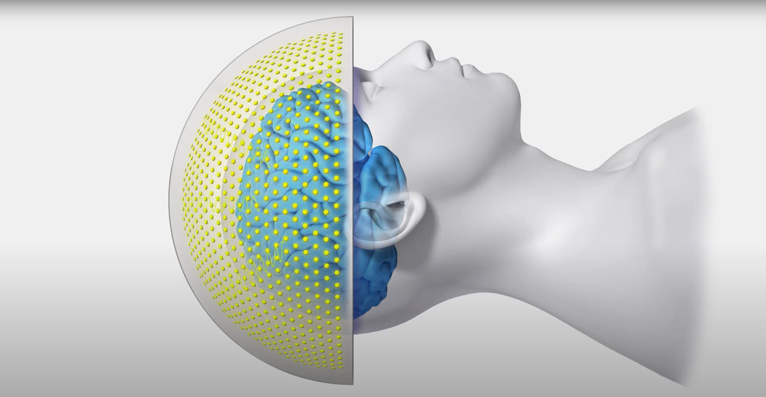
At Stanford Health Care (formerly Stanford Hospital & Clinics), experts in MR guided high intensity focused ultrasound (MRgFUS or MRgHIFU) now offer this non-invasive treatment as an alternative to more invasive RF ablation or surgical treatments of osteoid osteoma. The Food and Drug Administration has approved MRgFUS for the treatment of uterine fibroids and of painful bone metastases. Using the same technique as for the treatment of malignant bone cancers, MRgFUS has also been used as a non-invasive method to treat osteoid osteomas. MRgFUS treatment is performed under general, spinal or regional anesthesia in an outpatient setting, allowing the patient to return home in the afternoon after treatment. Compared with radiofrequency ablation, pain relief after MRgFUS is more rapid, occurring within the first day after the procedure. Also, because of the targeted nature of the focused ultrasound therapy, surrounding tissue is undamaged, thus allowing patients to return to normal activities within a week after the treatment.
With MRgFUS treatment, MR imaging is used to identify the nidus of the osteoid osteoma, followed by a few low energy ultrasound treatments that require a few minutes to heat the tumor and destroy it. MR imaging is then used to confirm the success of the treatment at the end of the procedure. The images below are from successful MRgFUS treatments at Stanford. The left image shows an osteoid osteoma in the left lower leg, in the tibia, and the right image demonstrates an osteoid osteoma in the right foot, in the fifth metatarsal. Patients' pain scores decreased to zero after treatment.
Complications
Risks associated with the ExAblate® procedure for osteoid osteoma are similar to those for treating bone metastasis, but are much less likely since the procedure generally involves one sonication with low energy.
Learn more about risks associated with treatment for bone metastasis and other potential short and long term complications of the ExAblate® procedure.